Research
Our active programs span the central and peripheral nervous systems and connect microscopy, image analysis, and quantitative biological readouts. In collaboration with bench scientists and clinicians, we develop reproducible imaging approaches that turn complex tissue data into measurable features of structure, cellular interaction, and disease biology.
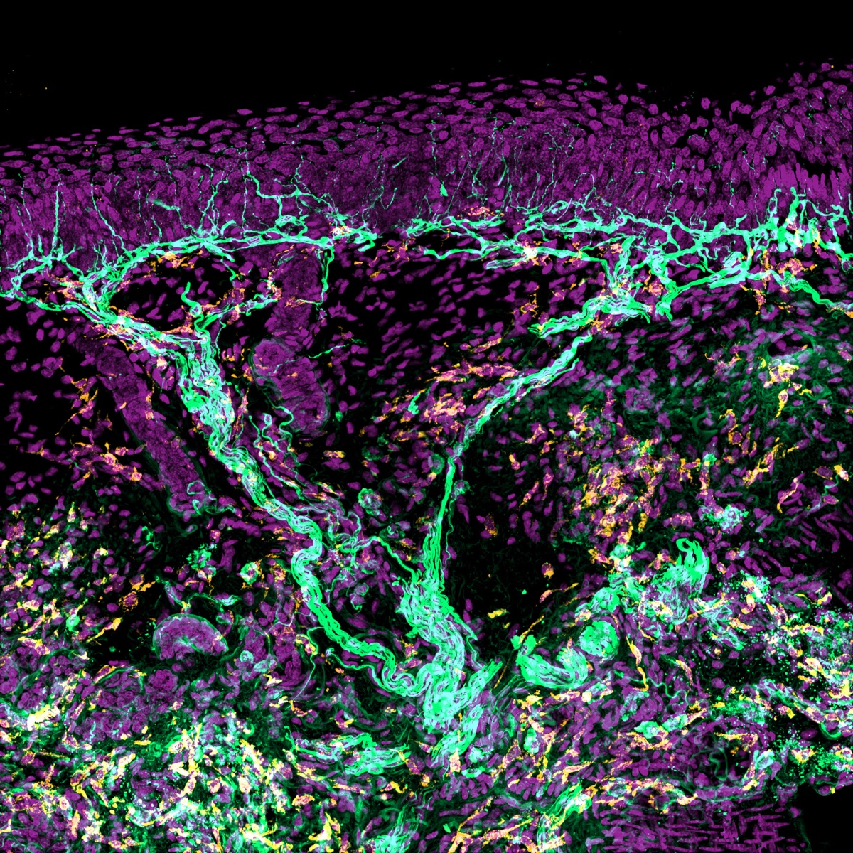
We study how sensory nerves, immune cells, lymphatics, vasculature, and skin cells interact during injury, regeneration, and chemotherapy-induced peripheral neuropathy. Quantitative analysis combines intraepidermal nerve fiber (IENF) density, S100A9 and other inflammatory readouts, and 3D reconstruction across skin biopsy, mouse hindpaw, and dorsal root ganglion assays. This work includes close collaboration with the Shin Lab and NIH-funded work with the Eisenmann Lab on damage-associated molecular patterns in chemotherapy-induced toxicity.
We study how axons, immune cells, and glial cells interact during regeneration, inflammatory injury, and demyelinating disease. The group develops quantitative frameworks for comparing immune cell distributions, tissue states, and regenerative outcomes across optic nerve, spinal cord, and experimental autoimmune encephalomyelitis (EAE) models. This work includes NIH-funded collaboration with the Jeon group at Johns Hopkins on immune-cell contributions to reinnervation after peripheral nerve injury.
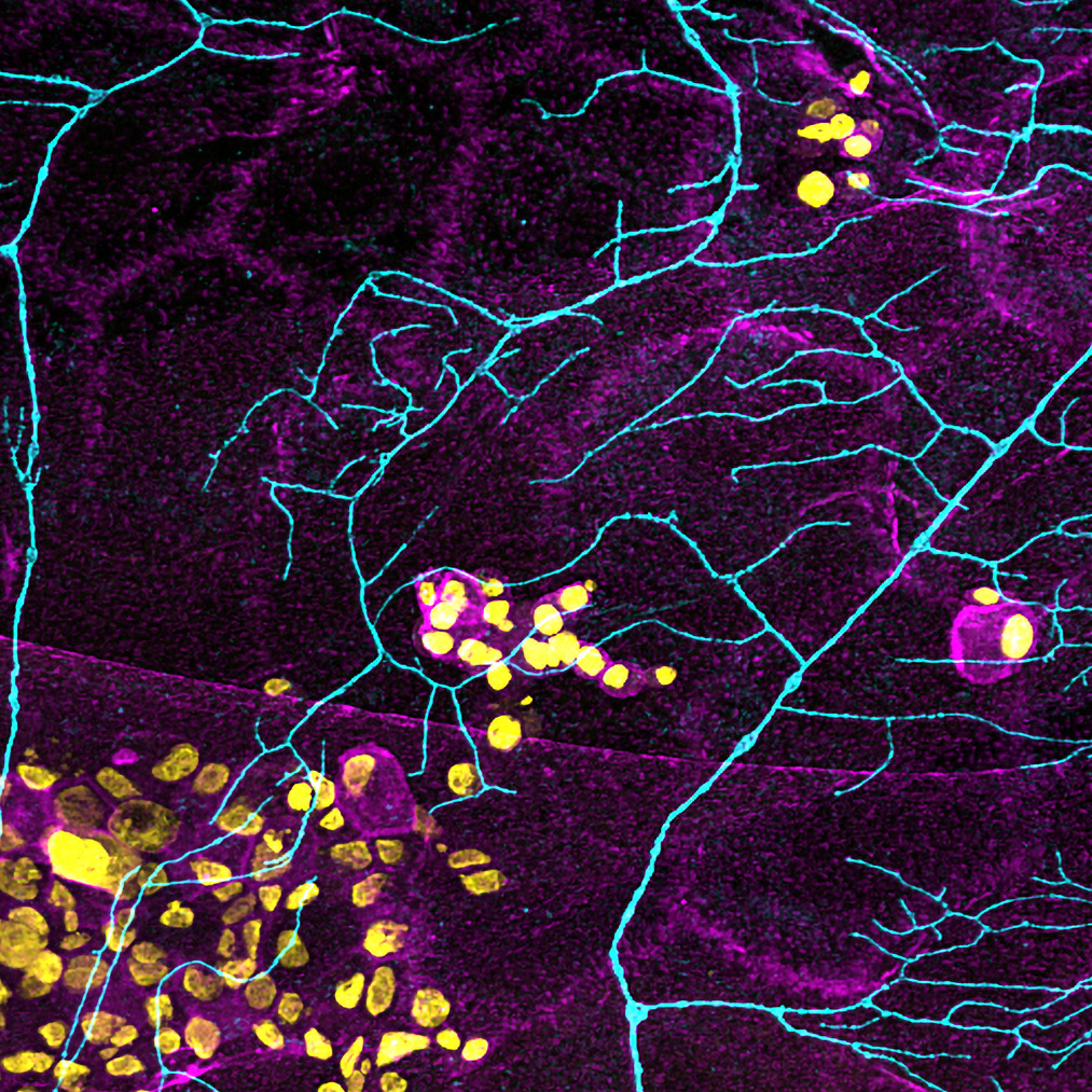
We develop improved tissue processing, clearing, imaging, and analysis methods for volumetric quantification of nerves, vasculature, immune cells, and lymphatics. This work supports reproducible 3D imaging and quantitative tissue analysis across intact and sectioned tissue preparations, with active applications in mouse hindpaw skin and human biopsy samples.
Quantitative tissue biology rarely belongs to one lab. Our active programs are built through sustained partnerships with collaborators at The Ohio State University and internationally, sharing tissue, reagents, methods, and analysis across groups so that biological expertise and quantitative imaging meet on the same problem.