
About the Quantitative
Imaging Group
The Quantitative Imaging Group in the Department of Neurology develops imaging and analysis frameworks that make microscopy a foundation for measurement, not just visualization. We build quantitative, auditable, and reproducible approaches for analyzing biological structure, cellular interactions, and tissue pathology across experimental and clinical research.
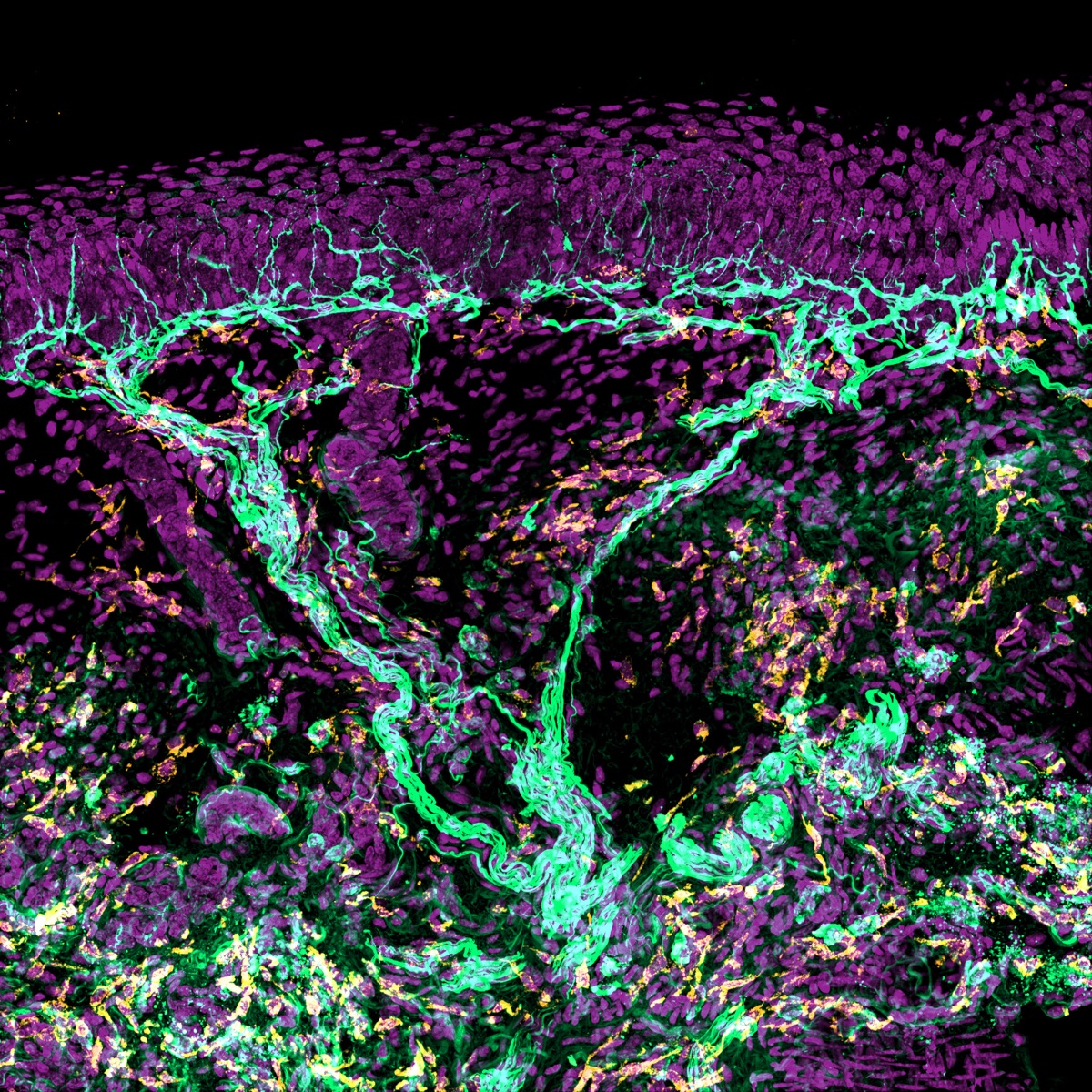
We study how sensory nerves, immune cells, lymphatics, vasculature, and skin cells interact during injury, regeneration, and chemotherapy-induced peripheral neuropathy. This work develops quantitative imaging approaches across human skin biopsy, mouse hindpaw, and dorsal root ganglion assays, in close collaboration with the Shin Lab.
We study how axons, immune cells, and glial cells interact during inflammatory injury, neurodegeneration, and regeneration in the central nervous system. This work develops quantitative imaging approaches across optic nerve, spinal cord, and experimental autoimmune encephalomyelitis models, in close collaboration with colleagues in the Department of Neurology.
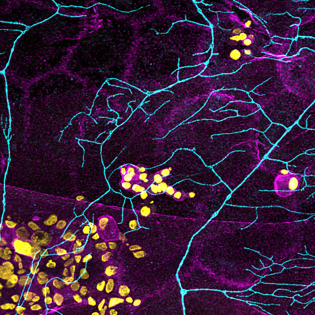
We develop improved fixation, tissue preparation, clearing, imaging, and analysis methods for volumetric quantification of nerves, vasculature, immune cells, and lymphatics. Active work spans mouse tissues and human biopsy samples.