NeuroMAP
Neurological Mapping and Phenotyping · The Ohio State University
NeuroMAP (Neurological Mapping and Phenotyping) is a transformative precision neuroscience initiative at The Ohio State University that integrates longitudinal clinical phenotyping with state-of-the-art molecular, imaging, and computational technologies to define the biological basis of neurological and psychiatric disease, discover biomarkers that predict disease progression and therapeutic response, and identify personalized therapeutic targets.
Participants are followed prospectively with serial blood and cerebrospinal fluid biomarkers, advanced neuroimaging, optical coherence tomography, neurological and neuropsychological assessments, and detailed clinical outcome measures, and may elect to donate tissues after death through NeuroCAPTURE, a rapid neuro-autopsy program that provides exceptionally high-quality central nervous system and peripheral tissues for research.
The defining feature of NeuroMAP is its ability to connect each participant's clinical journey during life with the molecular and cellular architecture of disease revealed after death, creating an unprecedented opportunity to determine how biomarkers, imaging findings, genetics, environmental exposures, and clinical progression reflect the underlying biology of human disease.

Five integrated discovery platforms
drive the program.
COMPASS tracks participants longitudinally, combining clinical and cognitive assessment, biospecimen collection, neuroimaging, retinal optical coherence tomography, and wearable-device monitoring into a single multimodal clinical phenotype.
NeuroCAPTURE is a rapid neuro-specific autopsy program that provides exceptionally high-quality tissue from the central nervous system and peripheral organs for research.
NeuroATLAS creates high-resolution three-dimensional cellular maps using tissue clearing, light-sheet microscopy, and custom machine and deep learning powered software.
NeuroCODEX performs comprehensive multimodal spatial molecular profiling to define the transcriptomic signatures and proteomic phenotype on a single cell level.
NeuroMODEL develops patient-derived iPSC and organoid models to validate biological discoveries and accelerate precision therapeutics.

Chair, Department of Neurology
Director, Neuroscience Research Institute
Co-director, Neurological Institute

Director, Clinical and Translational Research
Neurological Institute research ↗
Neuropathologist, Neuroscientist
Pathology profile ↗
Director, Quantitative Imaging
Quantitative Imaging ↗
Director, Neurobiology of Aging & Resilience Center
Director, Division of Neurogenetics

Associate Director, Neuroscience Research Institute
NRI Brain Bank ↗
Director, Center for Neuroimaging, Neurophenotyping, Neurocomputation and Neuromodulation (C4N)
C4N ↗
College of Optometry
Department of Ophthalmology & Visual Science

Vice Chair of Research,
Department of Biomedical Informatics

College of Optometry
Department of Ophthalmology & Visual Science
From the clinic to sub-cellular detail.
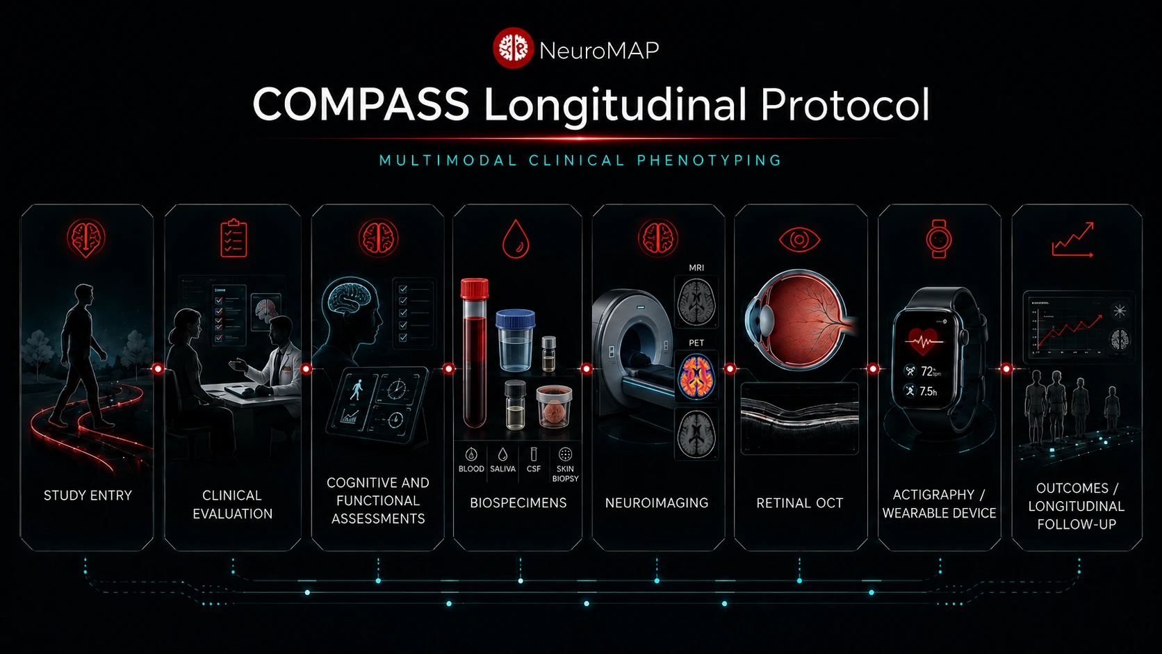
Conceptual plan for the NeuroMAP pipeline.

Conceptual plan for the NeuroMAP pipeline.
NeuroMAP extends beyond the brain and spinal cord to the interconnected systems that shape or reflect nervous system disease.
- Neuroimmune, gut-brain and peripheral systems: retina, meninges, cervical lymph nodes, skin, intestinal mucosa and other organs
- Across Alzheimer's disease, Parkinson's disease, ALS, multiple sclerosis, schizophrenia and related conditions
- AI integrates clinical course, genetics, environmental exposures, imaging and biomarkers from life with the spatial, cellular and molecular architecture revealed after death
- The blueprint spans the entire course of illness, from the earliest biological changes to end-stage pathology

Conceptual plan for the NeuroMAP pipeline.

Conceptual plan for the NeuroMAP pipeline.
Conceptual plan for the NeuroMAP pipeline.
Brain model derived from MRI by Nevit Dilmen, NIH 3D 3DPX-002739, used under CC BY 4.0.

Oscar Harari and Ayslan Castro Brant, 2026.
Deep-learning enabled 3D tissue analysis.
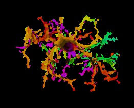
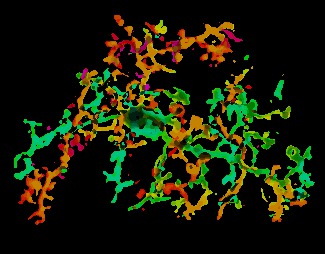
Animation shows optical sections at different depths revealing microglia populating the brain tissue and in proximity to amyloid plaques. Panels on the right show individually isolated microglia, with color representing depth.
Luke Hammond (Dept. Neurology) and Jie Gao (Dept. Neuroscience), 2024.
A digital blueprint of each disease.
Distinctively, NeuroMAP extends beyond the brain and spinal cord to investigate interconnected neuroimmune, gut-brain, and peripheral organ systems—including the retina, meninges, cervical lymph nodes, skin, intestinal mucosa, and other organs that shape or reflect nervous system disease.
Across disorders such as Alzheimer's disease, Parkinson's disease, amyotrophic lateral sclerosis, multiple sclerosis, schizophrenia, and related conditions, artificial intelligence will integrate every layer of information—from clinical course, genetics, environmental exposures, imaging, and biomarkers collected during life to the spatial, cellular, and molecular architecture revealed after death—creating an integrated digital blueprint of each disease that spans the entire course of illness—from the earliest biological changes to end-stage pathology.
By seamlessly linking clinical trajectories with the cellular and molecular architecture of disease, NeuroMAP will redefine our understanding of disease heterogeneity, reveal biomarkers that predict pathology, progression, and treatment response, uncover novel therapeutic targets, and establish a new paradigm for precision neuroscience that transforms the diagnosis, prevention, and treatment of neurological and psychiatric disease.